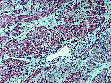
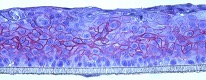
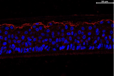
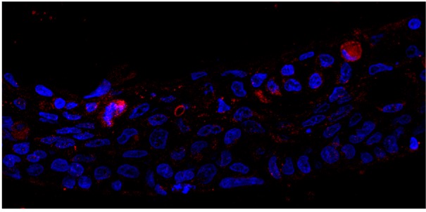
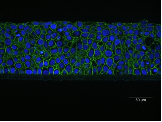
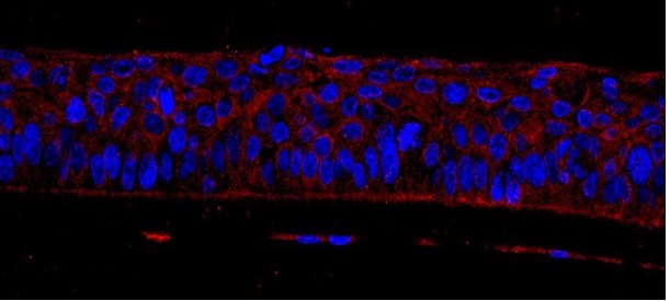
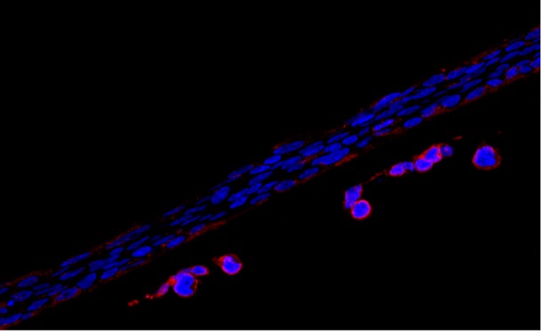
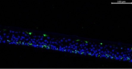
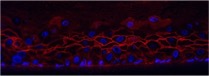
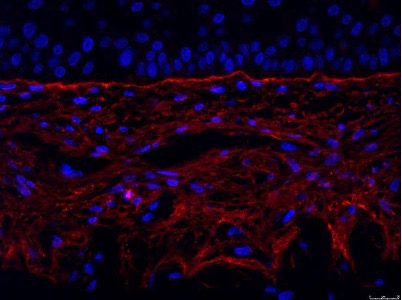
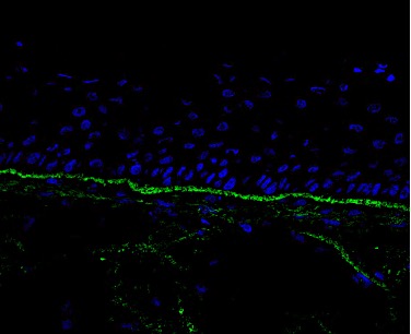
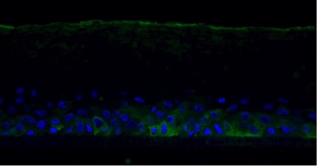
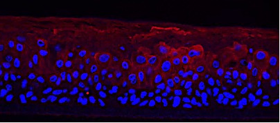
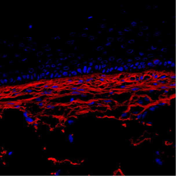
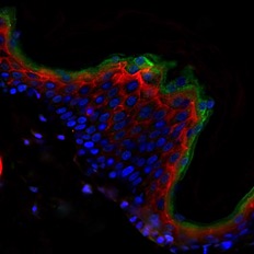
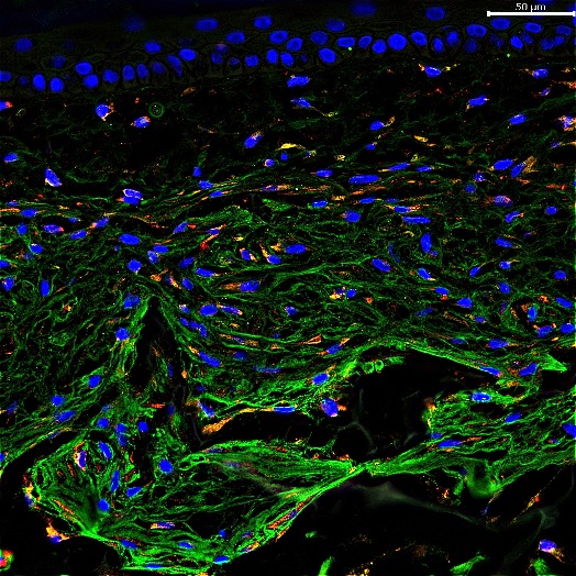
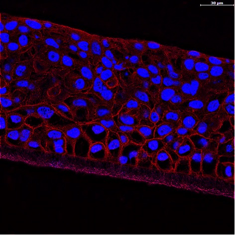
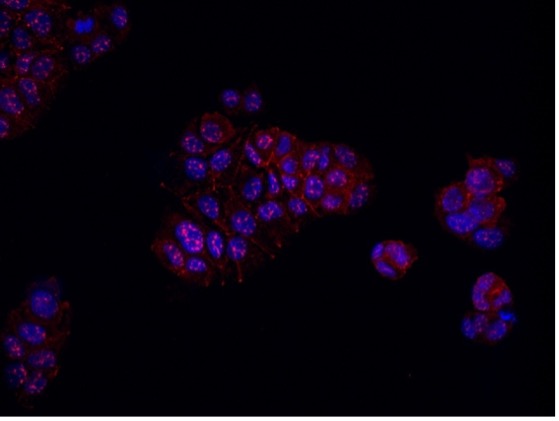
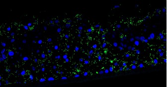
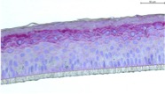
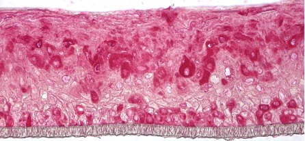
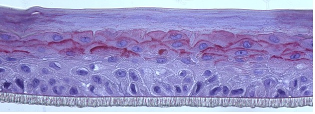
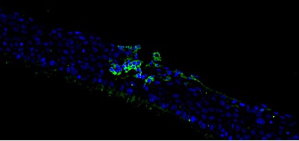
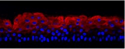
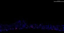
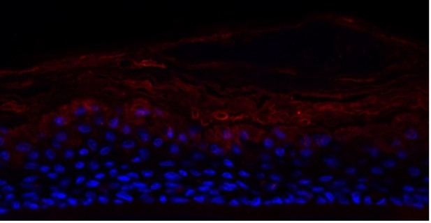
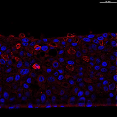
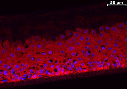
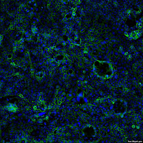
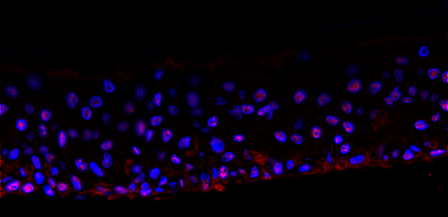
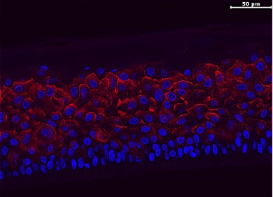
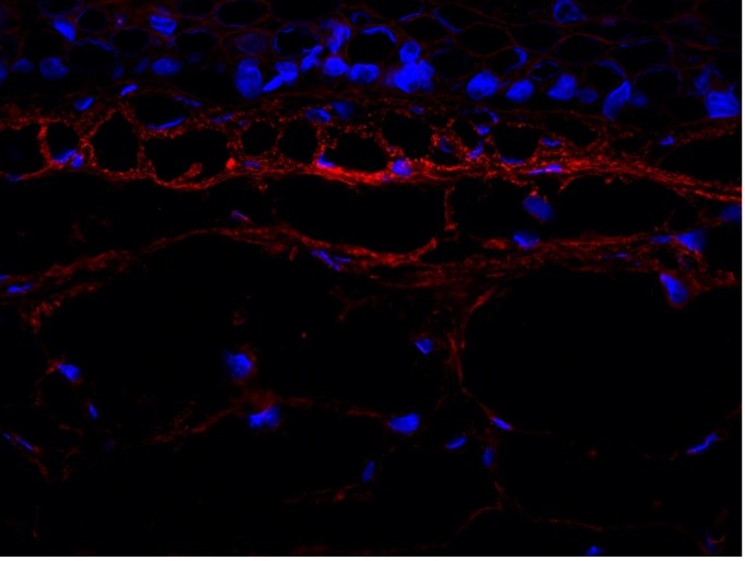
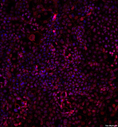
Histo-Morphology Platform2020-10-27T15:13:11+00:00

Evolution of VitroScreen’s histo-morphology platform

Since 2001 VitroScreen has been proposing the mechanism based Multiple Endpoint Analysis (MEA) approach on 3D human reconstructed tissues and scaffold free spheroids. Within MEA, morphological analysis is fundamental.

In 2017 VitroScreen enlarged its facilities strengthening its histological platform through additional capabilities and know-how: we perform standard and customized stainings, immunohistochemistry (IHC) and immunofluorescence (IF) on 3D tissue models, skin explants and spheroids.

VitroScreen has become in 2020 the first Italian CRO to offer services by Leica THUNDER Imager System – a new class of instruments designed specifically for high-quality, fast imaging of thick 3D specimens under optimal physiological conditions.

Leica THUNDER Imager is based on a fully motorized DMi8 microscope, Scanning Stage, highly sensitive camera, and multi-line, high-intensity fluorescence LED light source.

THUNDER Imagers remove the out-of-focus blur, that comes with three dimensional samples through Computational Clearing, an exclusive breakthrough technology from Leica Microsystems.

VitroScreen’s validated staining and biomarkers by IHC and IF

IHC/IF
STAINING

Applications

New 3D high content imaging platform

Leica THUNDER Imager by Leica provides an advanced solution for analysis on 3D cell cultures, such as spheroids and organoids.

Advantages and Unicity: high performance acquisition thanks to Leica Computational Clearing technology allowing to efficiently eliminate the signal components coming from the out of focus planes in real time, making it possible to study 3D samples with widefield fluorescence microscopes based on LED as light source.

The Computational Clearing, a new opto-digital imaging algorithm developed by Leica, is able to eliminate out-of-focus blur, differentiating between fluorescence signal and background by taking the size of the targeted specimen features into account. This innovative approach makes image details visible with high resolution if compared to the traditional light microscopy.

This innovative imaging approach allows the acquisition and processing of 3D whole mount samples, Z-stacks movie generation in high resolution, and 3D rendering simulations.